Nerve system
Sea Urchins with the lack of a true brain have a relatively simple nervous system, with radial nerves located on the inside of the test. Tube feet are the main source of sensory cells in the urchin with chemosensory and proposed light sensitivity through these multifunctional organs.
Water Vascular system
T. gratilla like all echinoderms have the the phylum unique system called the water vascular system. This system is composed of coelmic canals which are connected to the tube feet small outpockets which is powered by hydraulic pressure. This allows for movement by forcing the water into the tube feet allowing them to extend and relaxing of the musculature. The madeporite a canal that descends from the oboral opening of the urchin that descends and joins to the ring canal which encircles the esophagus. Radial canals extend from these ring canals and run along the outer skeleton of the urchin knowns as the test. The water vascular system in echinoderms is used by urchins for locomotion, feeding, internal transportation and respiration.
 Figure 2: T. gratilla extending tube feet that are placed under water as they are unable
to extend them without water. Photo: Stephanie Lyon
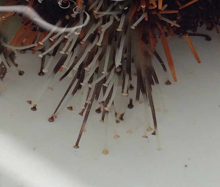 Figure 3: Tube feet of T. gratilla extended using the water vascular system squeezing
and releasing the bulb for extension and retraction. Photo: Stephanie Lyon
Urchins use this water vascular system to assist in the excretion of waste. The food enters the mouth via the use of Aristotle's lantern and the beak like structure. The teeth chew the food as the buccal cavity secrets a mucus that bins the particles into a pellet. The pellets then move through the body and enter the pharynx, which joins to the esophagus. The pellet then moves along the esophagus found on the outer side of the lantern before entering the widened stomach. From the stomach the pellet is broken down before moving to the intestine system which eventually leads to the rectum and the anus and the waste is expelled.
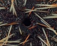 Figure 4:Oboral opening where waste is expelled
out the anus and into the water column.
Sea Urchins including
T. gratilla have a unique external skeleton structure know as the test. The test is made up of calcium carbonate and is used as a means of protection of the internal organs as well as maintaining the structure of the Urchin (Rupper et al 2004). Complimenting the test are short spines which a moveable and assists the urchin in movement and defense, these too are made out of similar material to the test (Ruppert et al 2004).
Information of these internal systems are summerised from Ruppert et al 2004 Chapter 28